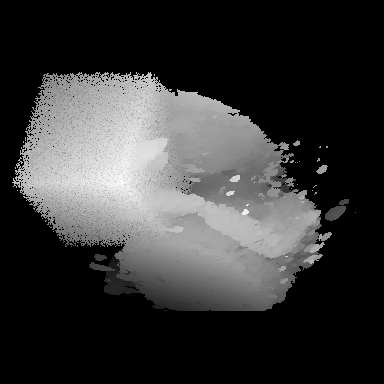

The results presented here are the results of experiments conducted using the Multiresolution Texture Segmenation (MTS) method. details of the research and experiments can be found in:
Muzzolini, R., Y.H. Yang, and R. Pierson, ``Three-dimensional segmentation of volume data,'' Proc. IEEE International Conference on Image Processing, Nov. 13-16, 1994, Austin, Texas, Vol. III, pp. 488-492.
Segmentation results for original ultrasound data of a human fetus. The volume data consists of 232 parallel slices from an in vitro linear scan of a fetus obtained using a 5-9 MHz ultrasound transducer. The inter-slice distance is 0.25 mm and the resolution of each image is 640 x 480 pixels (0.25 mm/pixel). A volume of size 256 x 256 x 128 was extracted from the original data and segmented. Results are shown using:
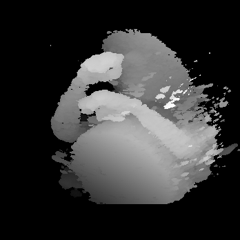
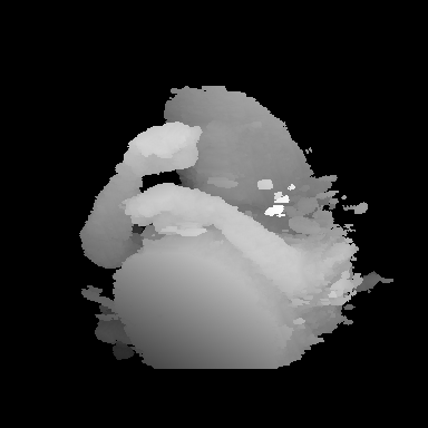
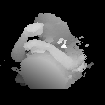
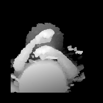
(e) Thresholding
