|
|
|
a)An original ultrasound image. |
b)After filtering. |
c)After edge detection. |
A long term project was started in 1990 in collaboration with Dr. R. Pierson of the Reproductive Research Unit at the Royal University Hospital of the University of Saskatchewan in applying and developing computer vision techniques in solving problems related to ultrasound imaging. Two new techniques in analysing ultrasound images have recently been completed. The first is a feature classification technique which can determine the best feature for classifying textures at various resolutions. The second technique, called Multiresolution Texture Segmentation, is proposed to perform segmentation of texture images. The experimental results on these techniques are very encouraging.
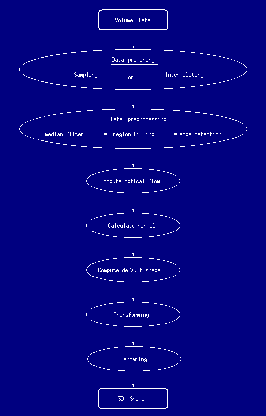
Recently, we have developed a rendering system using optical flow. The rendering system consists 7 parts, data preparing, data preprocessing, optical flow calculation, computing surface normal, computing default shape, transforming and rendering. Usually the original vloume data sets are not isotropic, so resampling or interpolation need to be done. The concentration of this work is on the medical ultrasound data visualization, since ultrasound is a widely used modality nowadays. Noise is the most serious problem in ultrasound image. In data preprocessing step, a median filter is applied to suppress the noise in the original images. Then a region analysis procedure is developed to fill the object region and get the outer most boundary of it. A speckle filter based on region analysis is applied to eliminate some noise points.
|
|
|
a)An original ultrasound image. |
b)After filtering. |
c)After edge detection. |
In this 3D recovery method, contours of the object are chosen as the representation of the surface. Computer Tomograph (CT) technology provide the ability to get the cross section of an object. The sequence of the cross sections obtained from scanning from the end to the other end of an object contains the 3D shape information. The uniqueness of the new method is that it introduced the concept of motion in the 3D shape reconstruction, since the sequence of images is obtained while there is a relative motion between the object and the scanner. Optical flow, which is a very useful tool of motion analysis, is used to compute the normal of the surface points on the contours. Default shape method is implemented to recover the whole surface from the information of the sequence of contours. Now the complete 3D shape of an object is recovered and can be transformed in order to be viewed from any direction. Phong's shading model and a Z-buffer algorithm are applied to render the 3D shape.
The synthetic images as well as real image data of MRI, Ultrasound and X-Ray CT are employed to test the feasibility of the 3D reconstruction method. Part of the results are shown in following.
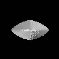
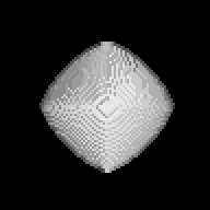
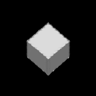
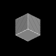
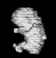
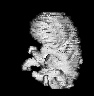
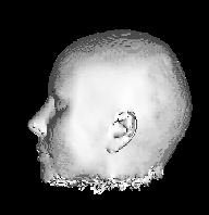
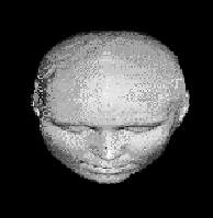
The whole system was written in C language on Sun SPARCstation 10 running SunOS 4.1.3. The following diagram depicts the whole system:
Some animation results are as follows: